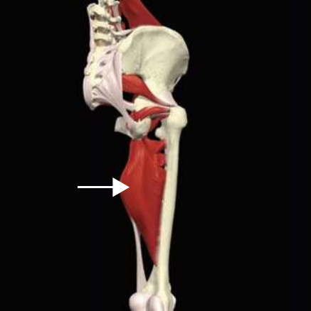
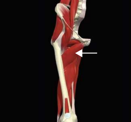
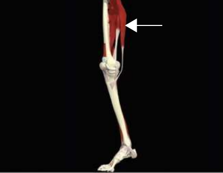
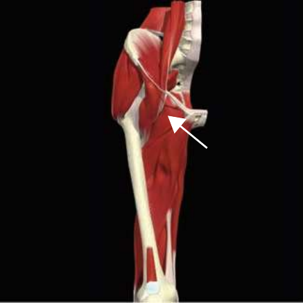
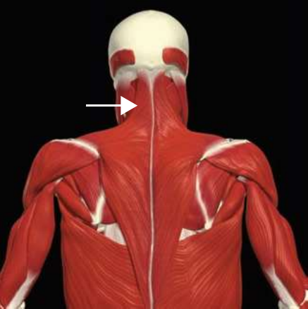
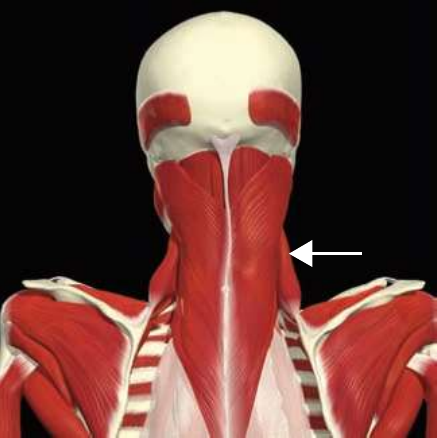
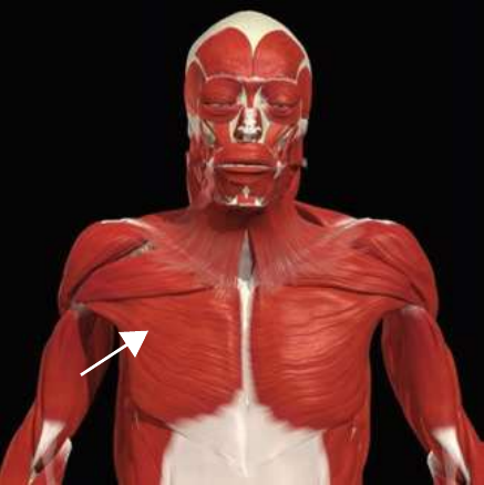
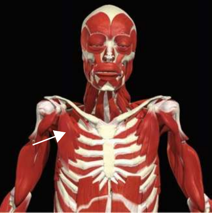
How the human movement system produces motion — planes & axes, joint actions, muscle roles and actions, leverage, and a full origin/insertion/function reference for every major muscle.
Adapted as a study guide from NASM's Introduction to Human Movement Science (filed here as ACE Ch‑17). Anatomical facts (origins, insertions, actions, innervation) reproduced for reference; teaching text authored for this guide. Figure slots are marked with their NASM captions for you to drop in licensed or original replacement art; the concept graphics and tables shown are original to this guide.
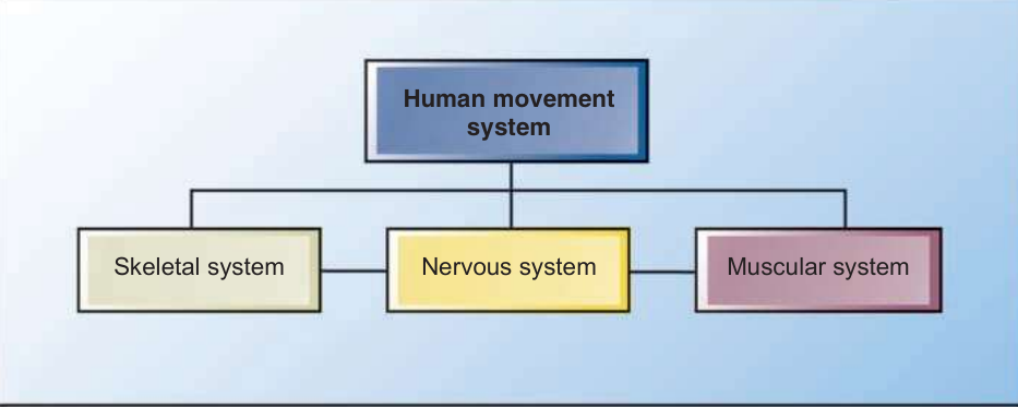
Human movement science is the study of how the human movement system (HMS) functions as one interdependent, interrelated scheme. It is built from three systems that look separate but must collaborate on every movement.
The muscular system supplies functional anatomy (the forces), the skeletal system supplies functional biomechanics (the levers and joints those forces act on), and the nervous system supplies motor behavior (the control and timing). Each system continuously gathers information about the body's relationship to its internal and external environment so it can produce the right movement pattern. When the three work together cleanly, you get optimum human movement; when one link is weak, the whole chain compensates.
Figure 2.1 — Components of the human movement system (figure slot; supply licensed or original art)
Biomechanics: a field that applies the principles of physics to quantitatively study how forces interact within a living body. This guide focuses on the motions the HMS produces (kinematics) and the forces acting on it (kinetics) — including anatomic terminology, planes of motion, joint motions, muscle action, force‑couples, leverage, and basic muscle mechanics.
Anatomic terminologyPlanes of Motion & Axes
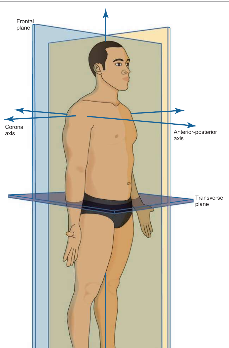
Human movement occurs in three dimensions and is described with a system of planes and axes. Three imaginary planes pass through the body at right angles, intersecting at the body's center of mass: the sagittal, frontal, and transverse planes. A movement is said to occur predominantly in a plane when it travels along or parallel to that plane — although no movement is ever strictly confined to one plane.
Movement in a plane rotates around an axis running perpendicular to it — much like the axle a car wheel revolves around. That rotation is what we call joint motion, and joint motions are named for their action in each plane.
Figure 2.2 — Planes of motion (figure slot; supply licensed or original art)
Plane → bisects → axis
Plane
Divides body into
Axis of rotation
Sagittal
Right & left halves
Frontal (coronal) axis
Frontal
Front & back halves
Anterior‑posterior axis
Transverse
Upper & lower halves
Longitudinal (vertical) axis
Plane 1The Sagittal Plane
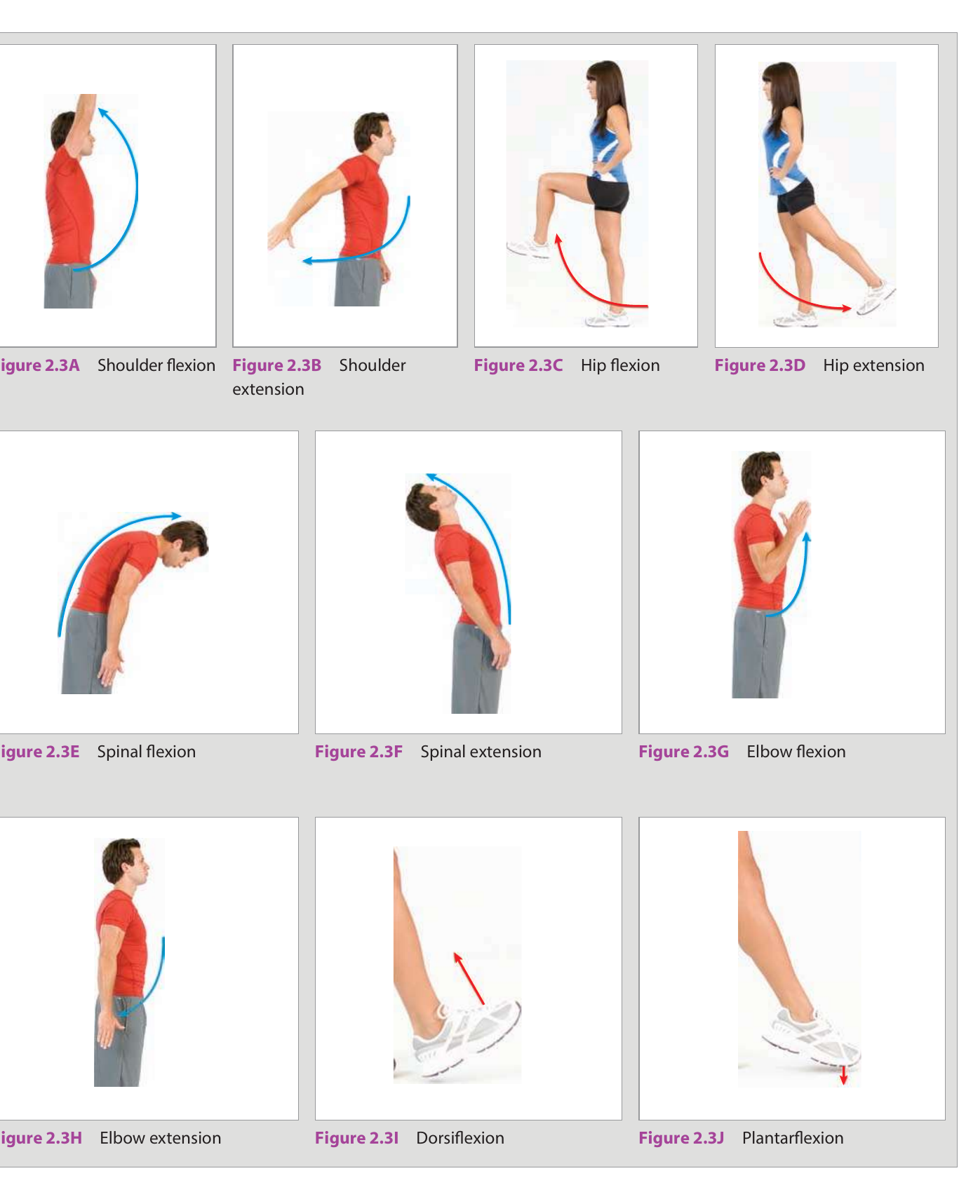
The sagittal plane bisects the body into right and left halves, and its motion occurs around a frontal axis. Its movements are flexion and extension. Flexion occurs when the relative angle between two adjacent segments decreases; extension occurs when that angle increases. These happen at many joints — vertebral, shoulder, elbow, wrist, hip, knee, foot, and hand.
The ankle is the exception, using special terms in the sagittal plane: "flexion" is more accurately called dorsiflexion and "extension" is called plantarflexion. Predominantly sagittal‑plane exercises include biceps curls, triceps pushdowns, squats, front lunges, calf raises, walking, running, and climbing stairs.
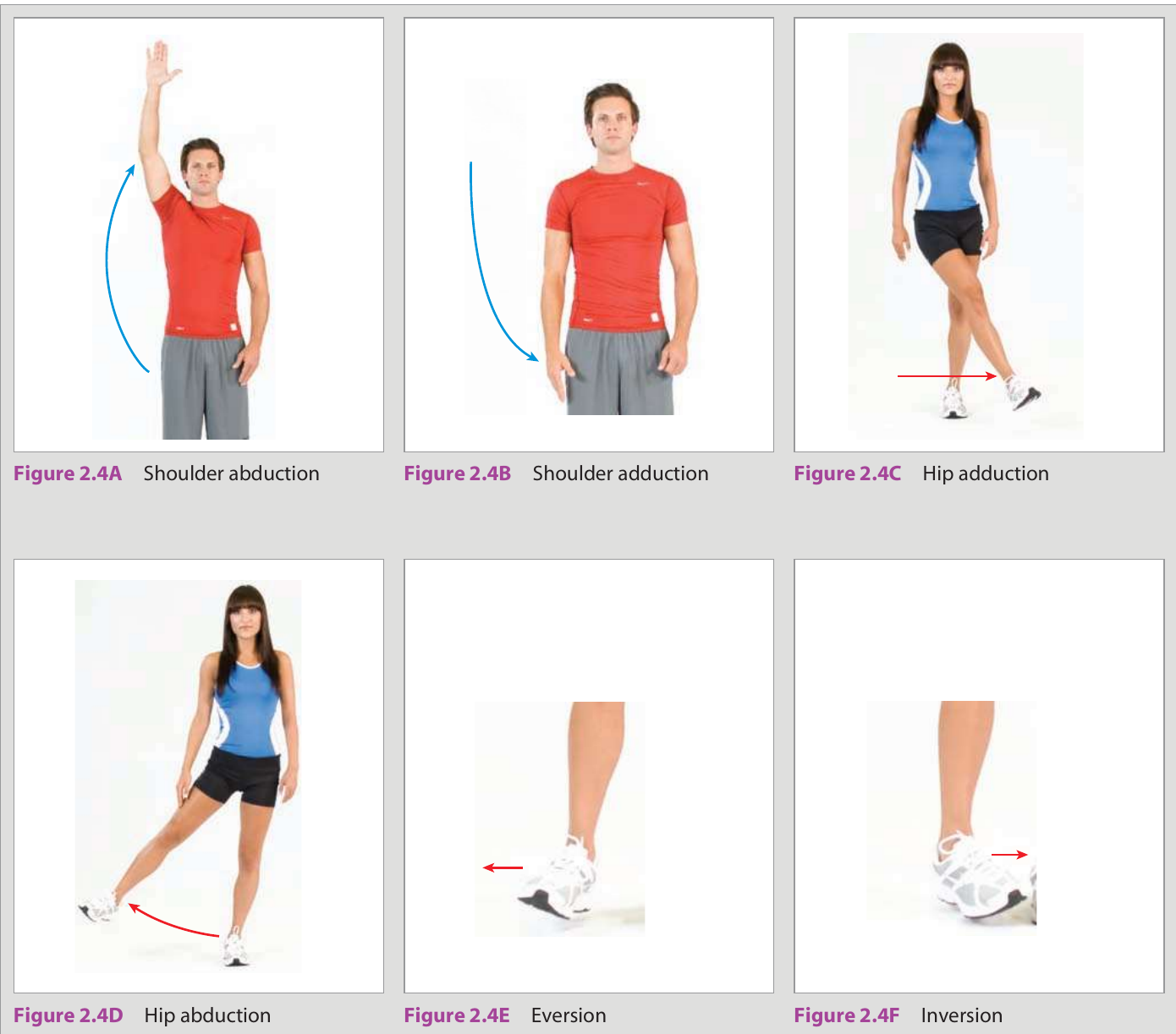
The frontal plane bisects the body into front and back halves, with motion occurring around an anterior‑posterior axis. Its movements are abduction and adduction of the limbs (relative to the trunk), lateral flexion of the spine, and eversion and inversion of the foot and ankle complex.
Abduction is movement away from the body's midline — an increase in the angle between two segments, but only in the frontal plane. Adduction is movement toward the midline — a decrease in that angle, again only in the frontal plane. Lateral flexion is side‑bending of the spine (cervical, thoracic, lumbar). Eversion and inversion specifically describe movement of the calcaneus and tarsals in the frontal plane during the functional movements of pronation and supination. Examples: lateral shoulder raises, side lunges, and side shuffling.
Figure 2.4 — Frontal-plane joint motions — shoulder & hip abduction/adduction; eversion & inversion (figure slot; supply licensed or original art)
Plane 3The Transverse Plane
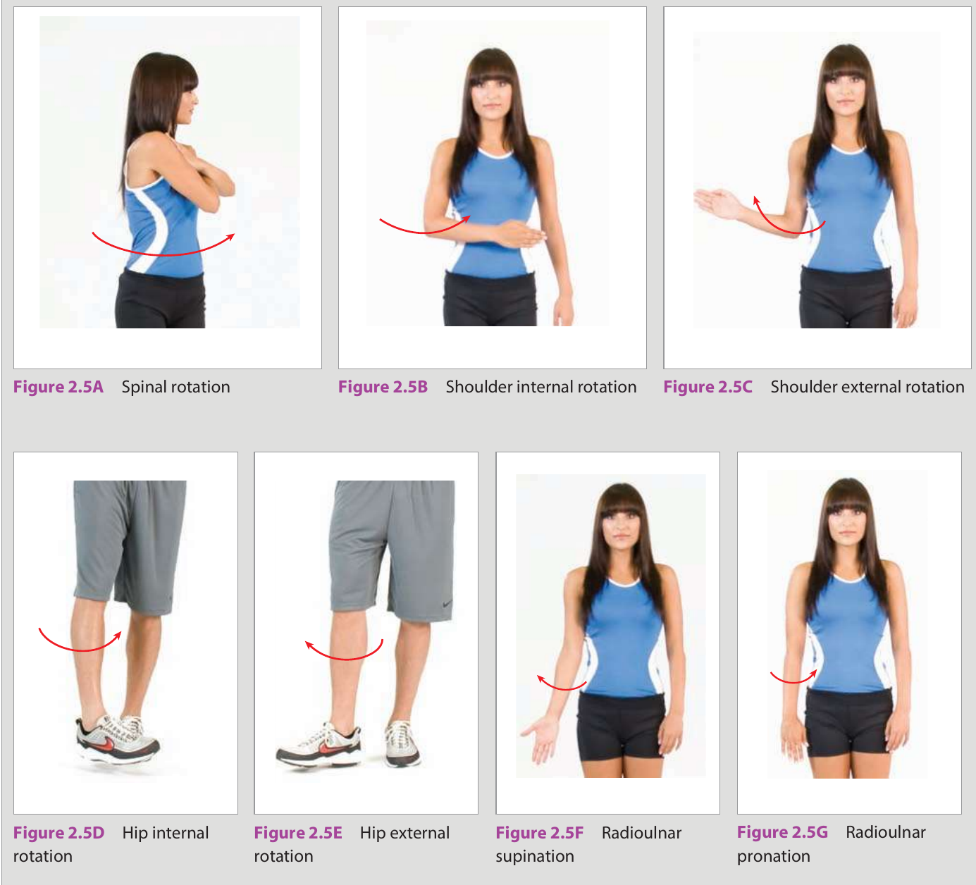
The transverse plane bisects the body into upper and lower halves, with motion occurring around a longitudinal (vertical) axis. Its movements are internal and external rotation of the limbs, right and left rotation of the head and trunk, and radioulnar pronation and supination of the forearm.
At the foot, transverse‑plane motions are termed abduction (toes pointing outward, externally rotated) and adduction (toes pointing inward, internally rotated). Examples of transverse‑plane training include cable rotations, turning lunges, throwing a ball, and swinging a bat.
Figure 2.5 — Transverse-plane joint motions — spinal rotation; shoulder & hip internal/external rotation; radioulnar pronation & supination (figure slot; supply licensed or original art)
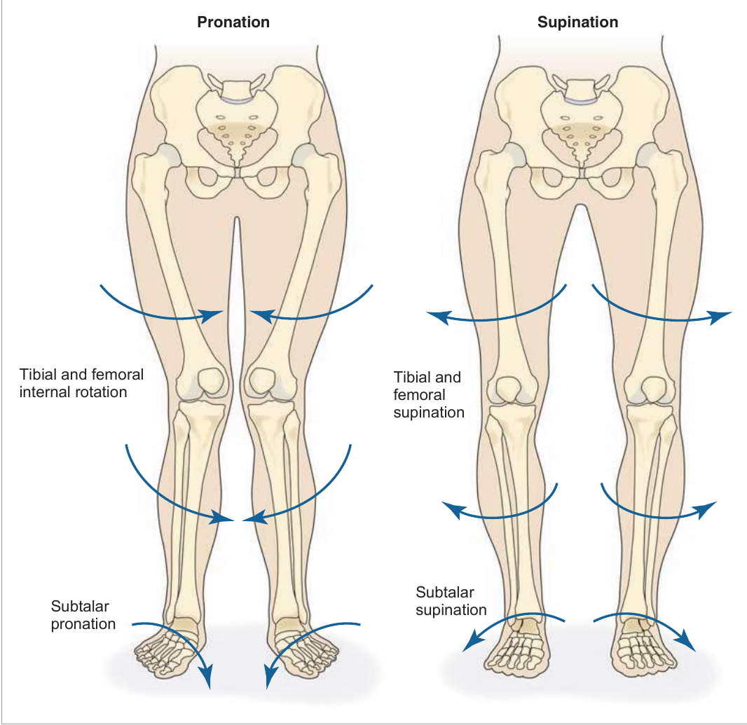
During real movement, the body must keep its center of gravity aligned over a constantly changing base of support. If alignment changes at one joint, other joints must change to compensate. Stand up and turn your kneecap inward, then outward, and you'll feel obligatory effects travel all the way from the subtalar joint to the pelvis.
When the patella turns inward (tibial and femoral internal rotation), pronation occurs at the subtalar joint. When it turns outward (tibial and femoral external rotation), subtalar supination occurs. Even though each joint has a predominant plane, every freely moveable joint shows some motion in all three planes.
This is the key insight: subtalar pronation, with obligatory tibial and femoral internal rotation, is a multiplanar, synchronized joint motion that occurs with eccentric muscle function (the body loading and decelerating). Subtalar supination, with obligatory tibial and femoral external rotation, is the matching multiplanar motion that occurs with concentric muscle function (the body producing force, as in push‑off).
Figure 2.6 — Lower extremity supination and pronation (figure slot; supply licensed or original art)
The gait cycle illustrates this interdependence. At initial contact, the subtalar joint pronates, creating obligatory internal rotation of the tibia, femur, and pelvis. At mid‑stance, the subtalar joint supinates, leading to obligatory external rotation up the chain. Remember the linkage is bidirectional: pelvic motion can create lower‑extremity motion and lower‑extremity motion can create pelvic motion.
Why it matters clinically. Poor control of subtalar pronation (with tibial/femoral internal rotation) reduces the ability to eccentrically decelerate multi‑segmental motion — a path to muscle imbalance, joint dysfunction, and injury. Poor production of subtalar supination (with external rotation) reduces the ability to concentrically produce push‑off force, which can lead to synergistic dominance.
ReferenceTable 2.2 — Functional Biomechanics
Because pronation and supination are tri‑planar, each joint of the lower extremity does three things at once during each. This is the cleanest summary of the chain:
During Pronation
Joint
Actions
Foot
Dorsiflexes, everts, abducts
Ankle
Dorsiflexes, everts, abducts
Knee
Flexes, adducts, internally rotates
Hip
Flexes, adducts, internally rotates
During Supination
Joint
Actions
Foot
Plantarflexes, inverts, adducts
Ankle
Plantarflexes, inverts, adducts
Knee
Extends, abducts, externally rotates
Hip
Extends, abducts, externally rotates
Muscle mechanicsMuscle Actions — Isolated vs Integrated Function
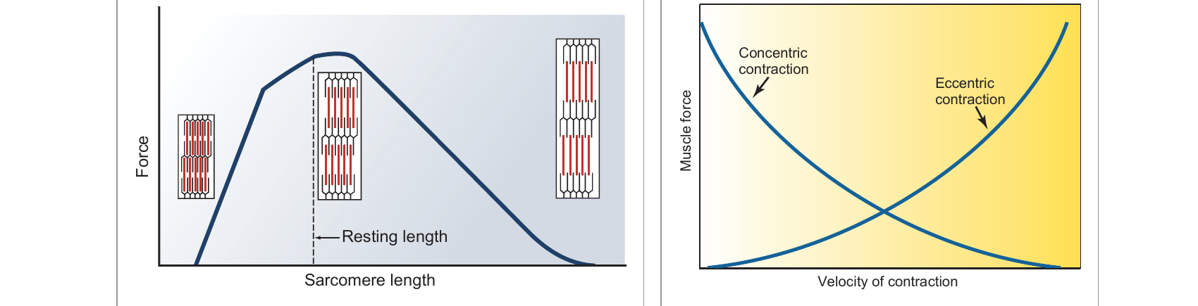
Every muscle in the reference below is described two ways, and understanding the difference is the whole game. Isolated function is what a muscle does when it shortens to accelerate a motion — its concentric action. Integrated function is what the same muscle does in real, multi‑joint movement, where it usually works eccentrically to decelerate the opposite motion, and isometrically to stabilize a joint while other muscles move.
The three muscle actions. In this guide's tables: Concentric = isolated function (accelerate), Eccentric = integrated function (decelerate), Isometric = integrated function (stabilize).
Reading the muscle tables. For each muscle you'll see its Origin, Insertion, Concentric action (what it accelerates), its Eccentric action (what it decelerates), its Isometric action (what it stabilizes), and its Innervation. Example — the gluteus maximus concentrically accelerates hip extension/external rotation, eccentrically decelerates hip flexion/internal rotation, and isometrically stabilizes the lumbo‑pelvic‑hip complex (LPHC).
Muscle mechanicsMuscle Roles in Movement
In any movement, muscles take one of four roles. Although their characteristics differ, all four work in concert to produce efficient motion.
Role
Definition
Example (hip extension)
Agonist
Muscle that acts as the prime mover
Gluteus maximus is the prime mover
Antagonist
Muscle that acts in direct opposition to the prime mover
Psoas (hip flexor) opposes the glute max
Synergist
Muscle that assists the prime mover during a movement pattern
Hamstring complex & erector spinae assist
Stabilizer
Muscle that supports/stabilizes the body while prime movers and synergists work
Transversus abdominis, internal oblique, multifidus, deep erector spinae stabilize the LPHC
The prime mover (agonist) is opposed by the antagonist, assisted by synergists, and supported by stabilizers that hold the LPHC steady.
Three mechanical concepts explain how well a muscle produces force.
Length‑tension relationship
A muscle generates its greatest force at its optimal (resting) length, where the actin and myosin filaments have the most available cross‑bridges. Stretch a sarcomere too long or shorten it too far and the available tension drops off — force falls on both sides of the optimum. Posture that holds a muscle chronically lengthened or shortened therefore weakens it.
Figure 2.10 — Length-tension relationships (figure slot; supply licensed or original art)
Force‑couple relationship
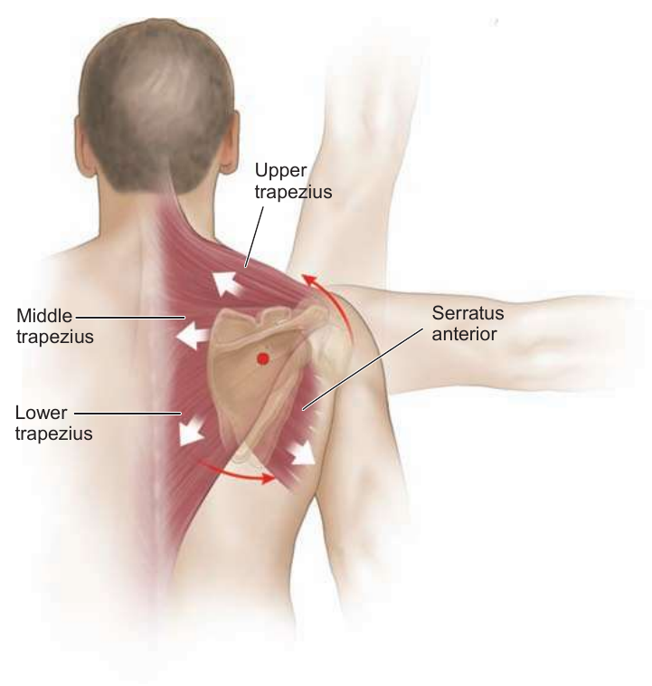
Muscles rarely act alone. A force‑couple is two or more muscles pulling in different directions to produce rotation around a joint — like two hands turning a steering wheel. The classic example is the scapula: the upper trapezius, lower trapezius, and serratus anterior pull in different lines yet together rotate the scapula upward so the arm can reach overhead. Normal length‑tension and proper force‑couples are what give a joint optimal neuromuscular control.
Figure 2.12 — Force-couple relationships (figure slot; supply licensed or original art)
Leverage & torque
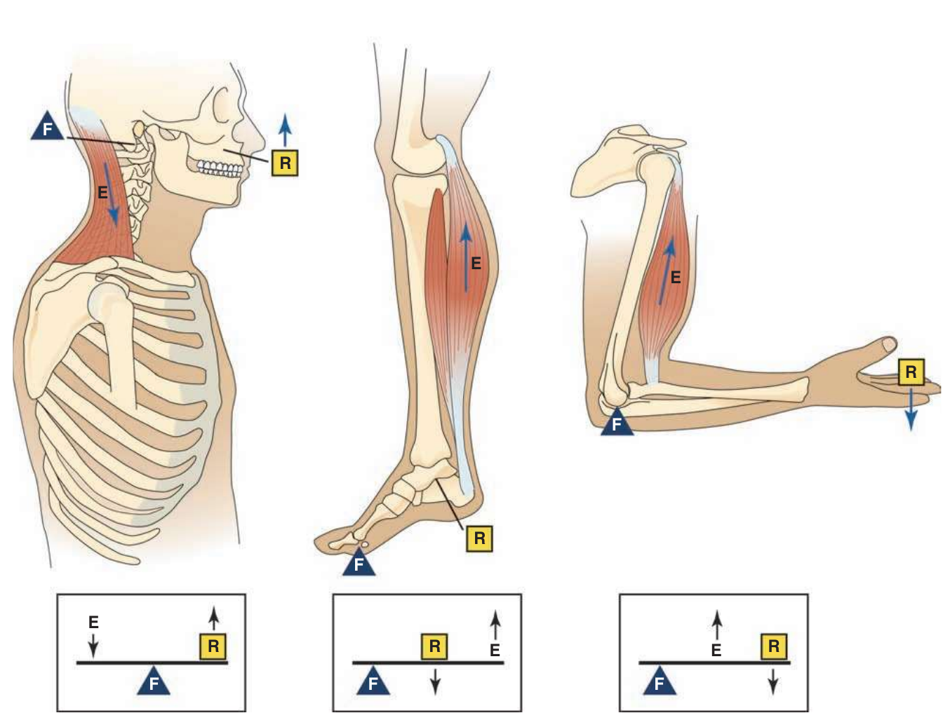
In the HMS, bones act as levers, joints act as fulcrums, muscles supply the force, and the body part (plus any external weight) is the load/resistance. Torque — the rotational force around a joint — equals force × distance from the fulcrum. There are three lever classes depending on the arrangement of fulcrum (F), effort/force (E), and load (L).
Figure 2.14 — Levers (figure slot; supply licensed or original art)
The big referenceMuscle Origin / Insertion / Function
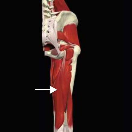
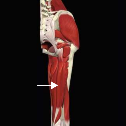
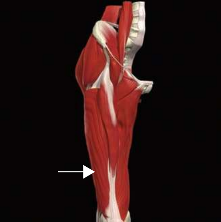
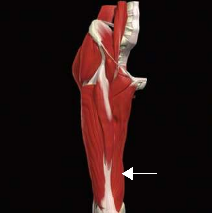
This is the core of the chapter — every major muscle with its origin, insertion, concentric (isolated) action, eccentric & isometric (integrated) actions, and innervation, organized by region. Each muscle is shown in the book's panel layout — a photo slot beside its full text block. Colour cues: Concentric · Eccentric · Isometric.
LEG COMPLEX
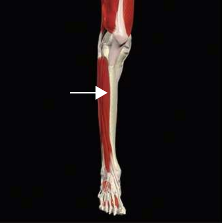
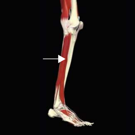
Anterior tibialis
Origin
Lateral condyle and proximal two-thirds of the lateral surface of the tibia
Insertion
Medial and plantar aspects of the medial cuneiform and the base of the first metatarsal
Isolated Function
Concentric Action
Ankle dorsiflexion and inversion
Integrated Function
Eccentric Action
Ankle plantar flexion and eversion
Isometric Action
Stabilizes the arch of the foot
Innervation
Deep peroneal nerve
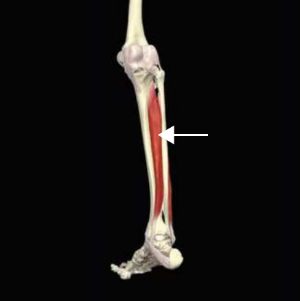
Posterior tibialis
Origin
Proximal two-thirds of posterior surface of the tibia and fibula
Insertion
Every tarsal bone (navicular, cuneiform, cuboid) but the talus, plus the bases of the 2nd–4th metatarsals; main insertion on the navicular tuberosity and medial cuneiform
Isolated Function
Concentric Action
Ankle plantar flexion and inversion of the foot
Integrated Function
Eccentric Action
Ankle dorsiflexion and eversion
Isometric Action
Stabilizes the arch of the foot
Innervation
Tibial nerve
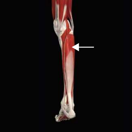
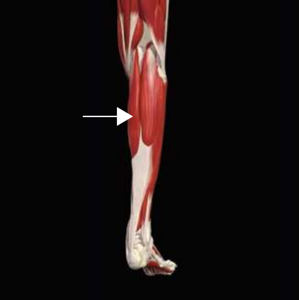
Soleus
Origin
Posterior surface of the fibular head and proximal one-third of its shaft, and the posterior side of the tibia
Insertion
Calcaneus via the Achilles tendon
Isolated Function
Concentric Action
Accelerates plantar flexion
Integrated Function
Eccentric Action
Decelerates ankle dorsiflexion
Isometric Action
Stabilizes the foot and ankle complex
Innervation
Tibial nerve
Gastrocnemius
Origin
Posterior aspect of the lateral and medial femoral condyles
Insertion
Calcaneus via the Achilles tendon
Isolated Function
Concentric Action
Accelerates plantar flexion
Integrated Function
Eccentric Action
Decelerates ankle dorsiflexion
Isometric Action
Isometrically stabilizes the foot and ankle complex
Innervation
Tibial nerve
Peroneus longus
Origin
Lateral condyle of tibia, head and proximal two-thirds of the lateral surface of the fibula
Insertion
Lateral surface of the medial cuneiform and lateral side of the base of the first metatarsal
Isolated Function
Concentric Action
Plantar flexes and everts the foot
Integrated Function
Eccentric Action
Decelerates ankle dorsiflexion and inversion
Isometric Action
Stabilizes the foot and ankle complex
Innervation
Superficial peroneal nerve
Biceps femoris – long head
Origin
Ischial tuberosity of the pelvis, part of the sacrotuberous ligament
Insertion
Head of the fibula
Isolated Function
Concentric Action
Accelerates knee flexion and hip extension, tibial external rotation
Integrated Function
Eccentric Action
Decelerates knee extension, hip flexion, and tibial internal rotation
Isometric Action
Stabilizes the lumbo-pelvic-hip complex and knee
Innervation
Tibial nerve
Biceps femoris – short head
Origin
Lower one-third of the posterior aspect of the femur
Insertion
Head of the fibula
Isolated Function
Concentric Action
Accelerates knee flexion and tibial external rotation
Integrated Function
Eccentric Action
Decelerates knee extension and tibial internal rotation
Isometric Action
Stabilizes the knee
Innervation
Common peroneal nerve
Semimembranosus
Origin
Ischial tuberosity of the pelvis
Insertion
Posterior aspect of the medial tibial condyle of the tibia
Isolated Function
Concentric Action
Accelerates knee flexion, hip extension and tibial internal rotation
Integrated Function
Eccentric Action
Decelerates knee extension, hip flexion and tibial external rotation
Isometric Action
Stabilizes the lumbo-pelvic-hip complex and knee
Innervation
Tibial nerve
Semitendinosus
Origin
Ischial tuberosity of the pelvis and part of the sacrotuberous ligament
Insertion
Proximal aspect of the medial tibial condyle of the tibia (pes anserine)
Isolated Function
Concentric Action
Accelerates knee flexion, hip extension and tibial internal rotation
Integrated Function
Eccentric Action
Decelerates knee extension, hip flexion and tibial external rotation
Isometric Action
Stabilizes the lumbo-pelvic-hip complex and knee
Innervation
Tibial nerve
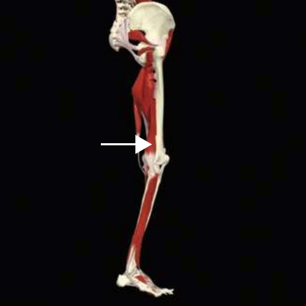
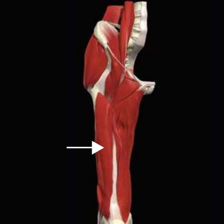
Vastus lateralis
Origin
Anterior and inferior border of the greater trochanter, lateral region of the gluteal tuberosity, lateral lip of the linea aspera of the femur
Insertion
Base of patella and tibial tuberosity of the tibia
Isolated Function
Concentric Action
Accelerates knee extension
Integrated Function
Eccentric Action
Decelerates knee flexion
Isometric Action
Stabilizes the knee
Innervation
Femoral nerve
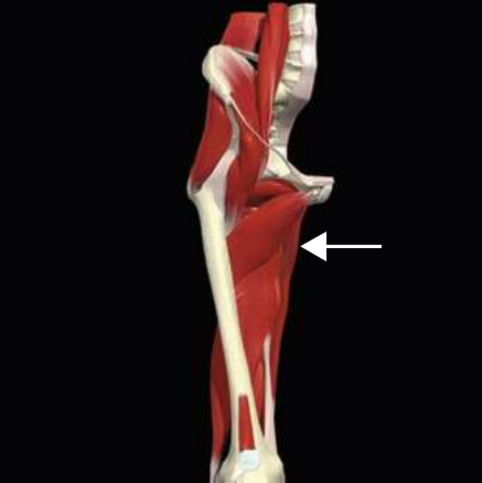
Vastus medialis
Origin
Lower region of intertrochanteric line, medial lip of linea aspera, proximal medial supracondylar line of the femur
Insertion
Base of patella, tibial tuberosity of the tibia
Isolated Function
Concentric Action
Accelerates knee extension
Integrated Function
Eccentric Action
Decelerates knee flexion
Isometric Action
Stabilizes the knee
Innervation
Femoral nerve
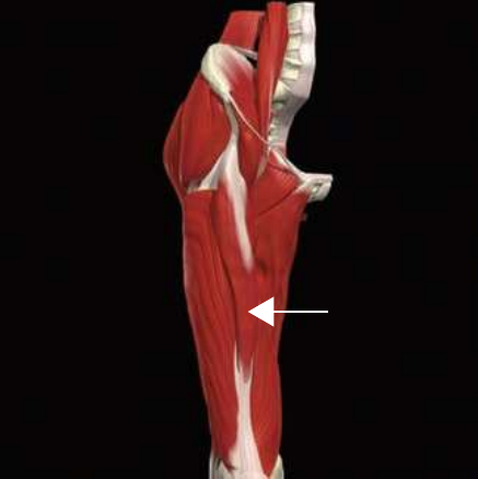
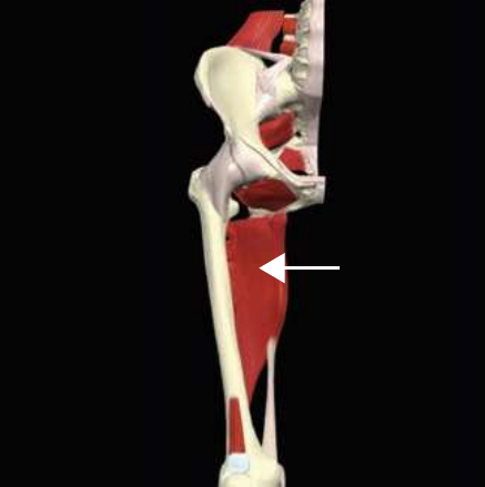
Vastus intermedius
Origin
Anterior-lateral regions of the upper two-thirds of the femur
Insertion
Base of patella, tibial tuberosity of the tibia
Isolated Function
Concentric Action
Accelerates knee extension
Integrated Function
Eccentric Action
Decelerates knee flexion
Isometric Action
Stabilizes the knee
Innervation
Femoral nerve
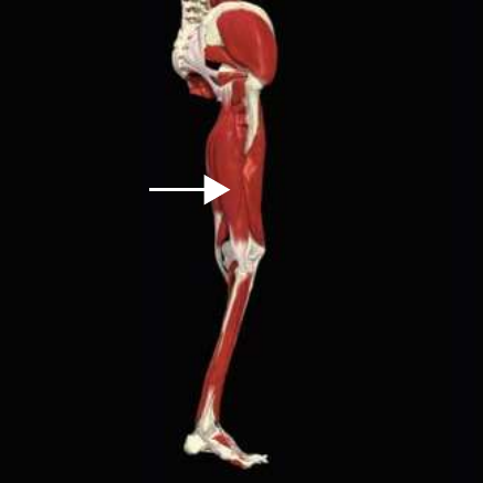
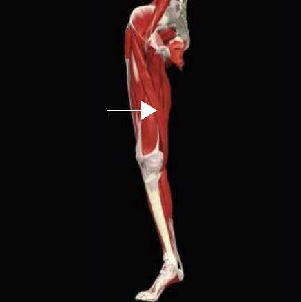
Rectus femoris
Origin
Anterior-inferior iliac spine of the pelvis
Insertion
Base of patella, tibial tuberosity of the tibia
Isolated Function
Concentric Action
Accelerates knee extension and hip flexion
Integrated Function
Eccentric Action
Decelerates knee flexion and hip extension
Isometric Action
Stabilizes the lumbo-pelvic-hip complex and knee
Innervation
Femoral nerve
HIP COMPLEX
Adductor longus
Origin
Anterior surface of the inferior pubic ramus of the pelvis
Insertion
Proximal one-third of the linea aspera of the femur
Isolated Function
Concentric Action
Accelerates hip adduction, flexion and internal rotation
Integrated Function
Eccentric Action
Decelerates hip abduction, extension and external rotation
Isometric Action
Stabilizes the lumbo-pelvic-hip complex
Innervation
Obturator nerve
Adductor magnus, anterior fibers
Origin
Ischial ramus of the pelvis
Insertion
Linea aspera of the femur
Isolated Function
Concentric Action
Accelerates hip adduction, flexion and internal rotation
Integrated Function
Eccentric Action
Decelerates hip abduction, extension and external rotation
Isometric Action
Stabilizes the lumbo-pelvic-hip complex
Innervation
Obturator nerve
Adductor magnus, posterior fibers
Origin
Ischial tuberosity of the pelvis
Insertion
Adductor tubercle on femur
Isolated Function
Concentric Action
Accelerates hip adduction, extension and external rotation
Integrated Function
Eccentric Action
Decelerates hip abduction, flexion and internal rotation
Isometric Action
Stabilizes the lumbo-pelvic-hip complex
Innervation
Sciatic nerve
Adductor brevis
Origin
Anterior surface of the inferior pubic ramus of the pelvis
Insertion
Proximal one-third of the linea aspera of the femur
Isolated Function
Concentric Action
Accelerates hip adduction, flexion and internal rotation
Integrated Function
Eccentric Action
Decelerates hip abduction, extension and external rotation
Isometric Action
Stabilizes the lumbo-pelvic-hip complex
Innervation
Obturator nerve
Gracilis
Origin
Anterior aspect of lower body of pubis
Insertion
Proximal medial surface of the tibia (pes anserine)
Isolated Function
Concentric Action
Accelerates hip adduction, flexion and internal rotation; assists in tibial internal rotation
Integrated Function
Eccentric Action
Decelerates hip abduction, extension and external rotation
Isometric Action
Stabilizes the lumbo-pelvic-hip complex and knee
Innervation
Obturator nerve
Pectineus
Origin
Pectineal line on the superior pubic ramus of the pelvis
Insertion
Pectineal line on the posterior surface of the upper femur
Isolated Function
Concentric Action
Accelerates hip adduction, flexion and internal rotation
Integrated Function
Eccentric Action
Decelerates hip abduction, extension and external rotation
Isometric Action
Stabilizes the lumbo-pelvic-hip complex
Innervation
Obturator nerve
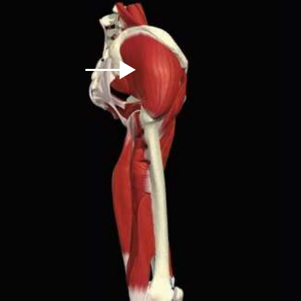
Gluteus medius, anterior fibers
Origin
Outer surface of the ilium
Insertion
Lateral surface of the greater trochanter on the femur
Isolated Function
Concentric Action
Accelerates hip abduction and internal rotation
Integrated Function
Eccentric Action
Decelerates hip adduction and external rotation
Isometric Action
Dynamically stabilizes the lumbo-pelvic-hip complex
Innervation
Superior gluteal nerve
Gluteus medius, posterior fibers
Origin
Outer surface of the ilium
Insertion
Lateral surface of the greater trochanter on the femur
Isolated Function
Concentric Action
Accelerates hip abduction and external rotation
Integrated Function
Eccentric Action
Decelerates hip adduction and internal rotation
Isometric Action
Stabilizes the lumbo-pelvic-hip complex
Innervation
Superior gluteal nerve
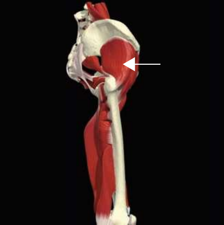
Gluteus minimus
Origin
Ilium between the anterior and inferior gluteal line
Insertion
Greater trochanter of the femur
Isolated Function
Concentric Action
Accelerates hip abduction, flexion, and internal rotation
Integrated Function
Eccentric Action
Decelerates frontal-plane hip adduction, extension, and external rotation
Isometric Action
Stabilizes the lumbo-pelvic-hip complex
Innervation
Superior gluteal nerve
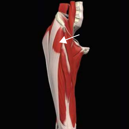
Tensor fascia latae
Origin
Outer surface of the iliac crest just posterior to the anterior-superior iliac spine of the pelvis
Insertion
Proximal one-third of the iliotibial band
Isolated Function
Concentric Action
Accelerates hip flexion, abduction and internal rotation
Integrated Function
Eccentric Action
Decelerates hip extension, adduction and external rotation
Isometric Action
Stabilizes the lumbo-pelvic-hip complex
Innervation
Superior gluteal nerve
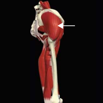
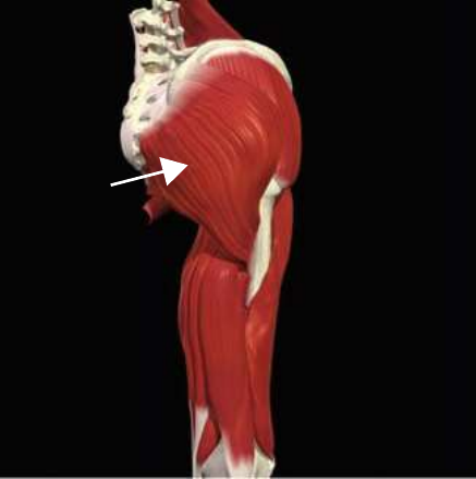
Gluteus maximus
Origin
Outer ilium, posterior side of sacrum and coccyx, and part of the sacrotuberous and posterior sacroiliac ligament
Insertion
Gluteal tuberosity of the femur and iliotibial tract
Isolated Function
Concentric Action
Accelerates hip extension and external rotation
Integrated Function
Eccentric Action
Decelerates hip flexion, internal rotation, and tibial internal rotation via the iliotibial band
Isometric Action
Stabilizes the lumbo-pelvic-hip complex
Innervation
Inferior gluteal nerve
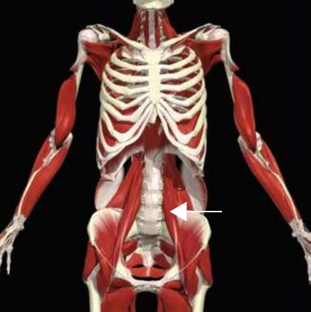
Psoas
Origin
Transverse processes and lateral bodies of the last thoracic and all lumbar vertebrae including intervertebral discs
Insertion
Lesser trochanter of the femur
Isolated Function
Concentric Action
Accelerates hip flexion and external rotation; extends and rotates the lumbar spine
Integrated Function
Eccentric Action
Decelerates hip internal rotation and decelerates hip extension
Isometric Action
Stabilizes the lumbo-pelvic-hip complex
Innervation
Spinal nerve branches of L2–L4
Sartorius
Origin
Anterior-superior iliac spine of the pelvis
Insertion
Proximal medial surface of the tibia
Isolated Function
Concentric Action
Accelerates hip flexion, external rotation and abduction; accelerates knee flexion and internal rotation
Integrated Function
Eccentric Action
Decelerates hip extension, external rotation, knee extension and external rotation
Isometric Action
Stabilizes the lumbo-pelvic-hip complex and knee
Innervation
Femoral nerve
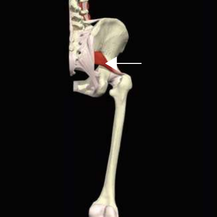
Piriformis
Origin
Anterior surface of the sacrum
Insertion
The greater trochanter of the femur
Isolated Function
Concentric Action
Accelerates hip external rotation, abduction and extension
Integrated Function
Eccentric Action
Decelerates hip internal rotation, adduction and flexion
Isometric Action
Stabilizes the hip and sacroiliac joints
Innervation
Sciatic nerve
ABDOMINAL MUSCULATURE
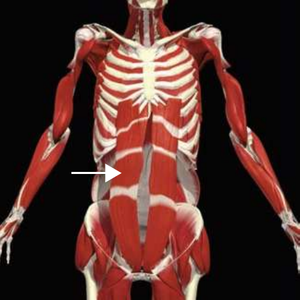
Rectus abdominis
Origin
Pubic symphysis of the pelvis
Insertion
Ribs 5–7
Isolated Function
Concentric Action
Spinal flexion, lateral flexion and rotation
Integrated Function
Eccentric Action
Spinal extension, lateral flexion and rotation
Isometric Action
Stabilizes the lumbo-pelvic-hip complex
Innervation
Intercostal nerve T7–T12
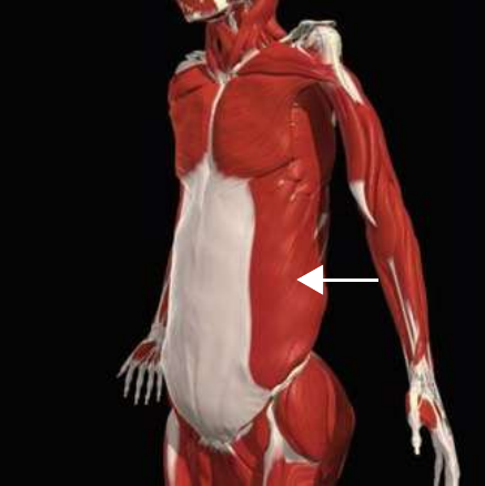
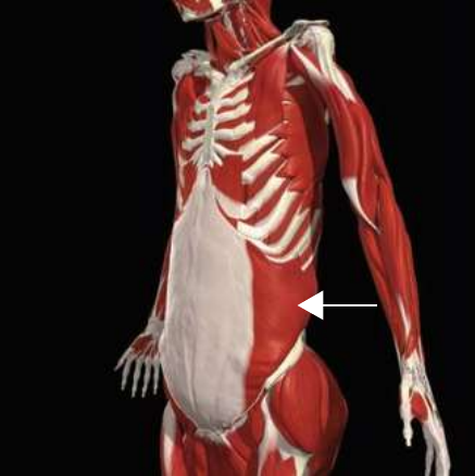
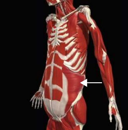
External oblique
Origin
External surface of ribs 4–12
Insertion
Anterior iliac crest of the pelvis, linea alba and contralateral rectus sheaths
Isolated Function
Concentric Action
Spinal flexion, lateral flexion and contralateral rotation
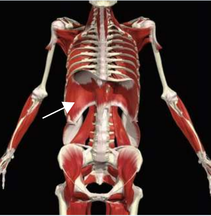
Costal part: inner surfaces of the cartilages and adjacent bony regions of ribs 6–12. Sternal part: posterior side of the xiphoid process. Crural (lumbar) part: two aponeurotic arches over the quadratus lumborum and psoas major, plus right and left crus from the bodies of L1–L3 and their intervertebral discs
Insertion
Central tendon
Isolated Function
Concentric Action
Pulls the central tendon inferiorly, increasing the volume in the thoracic cavity
Integrated Function
Isometric Action
Stabilization of the lumbo-pelvic-hip complex
Innervation
Phrenic nerve (C3–C5)
BACK MUSCULATURE
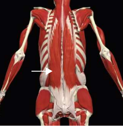
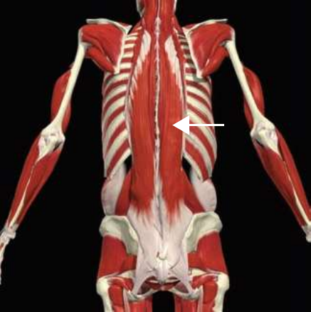
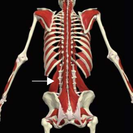
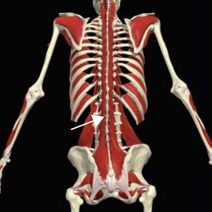
Iliocostalis: lumborum division
Origin
Common origin: iliac crest of the pelvis, sacrum, & the spinous & transverse processes of T1–L5
Insertion
Inferior border of ribs 7–12
Isolated Function
Concentric Action
Spinal extension, rotation and lateral flexion
Integrated Function
Eccentric Action
Spinal flexion, rotation and lateral flexion
Isometric Action
Stabilizes the spine during functional movements
Innervation
Dorsal rami of thoracic and lumbar nerves
Iliocostalis: thoracis division
Origin
Common origin: iliac crest of the pelvis, sacrum, & the spinous & transverse processes of T1–L5
Insertion
Superior border of ribs 1–6
Isolated Function
Concentric Action
Spinal extension, rotation and lateral flexion
Integrated Function
Eccentric Action
Spinal flexion, rotation and lateral flexion
Isometric Action
Stabilizes the spine during functional movements
Innervation
Dorsal rami of thoracic nerves
Iliocostalis: cervicus division
Origin
Common origin: iliac crest of the pelvis, sacrum, & the spinous & transverse processes of T1–L5
Insertion
Transverse process of C4–C6
Isolated Function
Concentric Action
Spinal extension, rotation and lateral flexion
Integrated Function
Eccentric Action
Spinal flexion, rotation and lateral flexion
Isometric Action
Stabilizes the spine during functional movements
Innervation
Dorsal rami of thoracic nerves
Longissimus: thoracis division
Origin
Common origin: iliac crest of the pelvis, sacrum, & the spinous & transverse processes of T1–L5
Insertion
Transverse process T1–T12; Ribs 2–12
Isolated Function
Concentric Action
Spinal extension, rotation and lateral flexion
Integrated Function
Eccentric Action
Spinal flexion, rotation and lateral flexion
Isometric Action
Stabilizes the spine during functional movements
Innervation
Dorsal rami of thoracic and lumbar nerves
Longissimus: cervicus division
Origin
Common origin: iliac crest of the pelvis, sacrum, & the spinous & transverse processes of T1–L5
Insertion
Transverse process of C6–C2
Isolated Function
Concentric Action
Spinal extension, rotation and lateral flexion
Integrated Function
Eccentric Action
Spinal flexion, rotation and lateral flexion
Isometric Action
Stabilizes the spine during functional movements
Innervation
Dorsal rami of cervical nerves
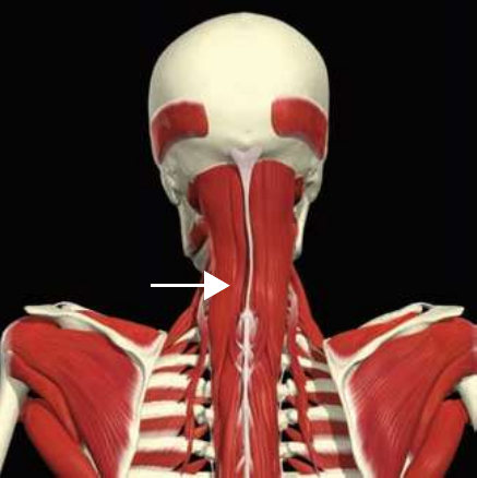
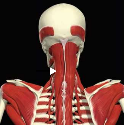
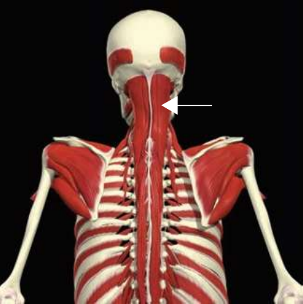
Longissimus: capitis division
Origin
Common origin: iliac crest of the pelvis, sacrum, & the spinous & transverse processes of T1–L5
Insertion
Mastoid process of the skull
Isolated Function
Concentric Action
Spinal extension, rotation and lateral flexion
Integrated Function
Eccentric Action
Spinal flexion, rotation and lateral flexion
Isometric Action
Stabilizes the spine during functional movements
Innervation
Dorsal rami of cervical nerves
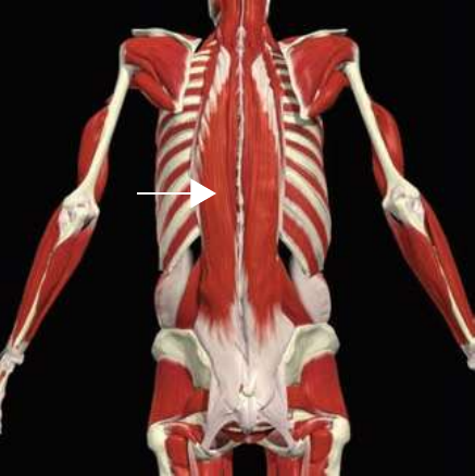
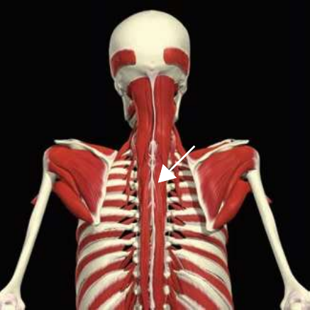
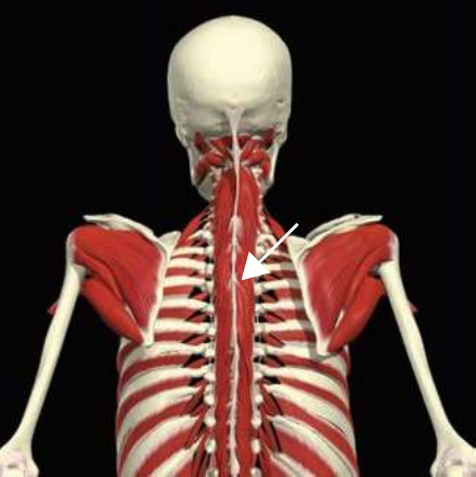
Spinalis: thoracis division
Origin
Common origin: iliac crest of the pelvis, sacrum, & the spinous & transverse processes of T1–L5
Insertion
Spinous process of T7–T4
Isolated Function
Concentric Action
Spinal extension, rotation and lateral flexion
Integrated Function
Eccentric Action
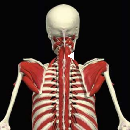
Spinal flexion, rotation and lateral flexion
Isometric Action
Stabilizes the spine during functional movements
Innervation
Dorsal rami of thoracic nerves
Spinalis: cervicus division
Origin
Common origin: iliac crest of the pelvis, sacrum, & the spinous & transverse processes of T1–L5
Insertion
Spinous process of C3–C2
Isolated Function
Concentric Action
Spinal extension, rotation and lateral flexion
Integrated Function
Eccentric Action
Spinal flexion, rotation and lateral flexion
Isometric Action
Stabilizes the spine during functional movements
Innervation
Dorsal rami of cervical nerves
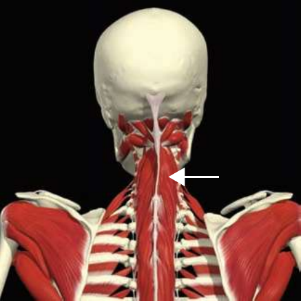
Spinalis: capitis division
Origin
Common origin: iliac crest of the pelvis, sacrum, & the spinous & transverse processes of T1–L5
Insertion
Between the superior and inferior nuchal lines on the occipital bone of the skull
Isolated Function
Concentric Action
Spinal extension, rotation and lateral flexion
Integrated Function
Eccentric Action
Spinal flexion, rotation and lateral flexion
Isometric Action
Stabilizes the spine during functional movements
Innervation
Dorsal rami of cervical nerves
Quadratus lumborum
Origin
Iliac crest of the pelvis
Insertion
12th rib, transverse processes L2–L5
Isolated Function
Concentric Action
Spinal lateral flexion
Integrated Function
Eccentric Action
Decelerates contralateral lateral spinal flexion
Isometric Action
Stabilizes the lumbo-pelvic-hip complex
Innervation
Spinal nerves (T12–L3)
Transversospinalis: thoracis division
Origin
Transverse process T12–T7
Insertion
Spinous process T4–C6
Isolated Function
Concentric Action
Produces spinal extension and lateral flexion; extension and contralateral rotation of the head
Integrated Function
Eccentric Action
Decelerates lateral flexion of the spine, flexion and contralateral rotation of the head
Isometric Action
Stabilizes the spine
Innervation
Dorsal rami C1–T6 spinal nerves
Transversospinalis: cervicis division
Origin
Transverse process T6–C4
Insertion
Spinous process C5–C2
Isolated Function
Concentric Action
Produces spinal extension and lateral flexion; extension and contralateral rotation of the head
Integrated Function
Eccentric Action
Decelerates lateral flexion of the spine, flexion and contralateral rotation of the head
Isometric Action
Stabilizes the spine
Innervation
Dorsal rami C1–T6 spinal nerves
Transversospinalis: capitus division
Origin
Transverse process T6–C7; Articular process C6–C4
Insertion
Nuchal line of the occipital bone of the skull
Isolated Function
Concentric Action
Produces spinal extension and lateral flexion; extension and contralateral rotation of the head
Integrated Function
Eccentric Action
Decelerates lateral flexion of the spine, flexion and contralateral rotation of the head
Isometric Action
Stabilizes the spine
Innervation
Dorsal rami C1–T6 spinal nerves
Multifidus
Origin
Posterior aspect of the sacrum; processes of the lumbar, thoracic and cervical spine
Insertion
Spinous processes 1 to 4 segments above the origin
Isolated Function
Concentric Action
Spinal extension and contralateral rotation
Integrated Function
Eccentric Action
Spinal flexion and rotation
Isometric Action
Stabilizes the spine
Innervation
Corresponding spinal nerves
SHOULDER MUSCULATURE
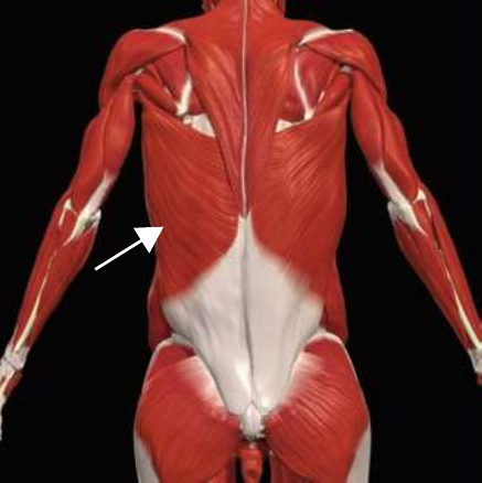
Latissimus dorsi
Origin
Spinous processes of T7–T12; iliac crest of the pelvis; thoracolumbar fascia; ribs 9–12
Insertion
Inferior angle of the scapula; intertubercular groove of the humerus
Isolated Function
Concentric Action
Shoulder extension, adduction and internal rotation
Integrated Function
Eccentric Action
Shoulder flexion, abduction and external rotation, and spinal flexion
Isometric Action
Stabilizes the lumbo-pelvic-hip complex and shoulder
Innervation
Thoracodorsal nerve (C6–C8)
Serratus anterior
Origin
Ribs 4–12
Insertion
Medial border of the scapula
Isolated Function
Concentric Action
Scapular protraction
Integrated Function
Eccentric Action
Scapular retraction
Isometric Action
Stabilizes the scapula
Innervation
Long thoracic nerve (C5–C7)
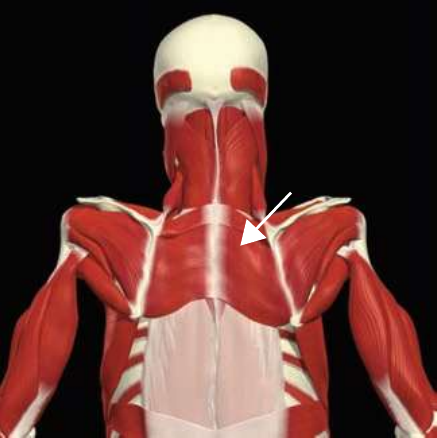
Rhomboids
Origin
Spinous processes of C7–T5
Insertion
Medial border of the scapula
Isolated Function
Concentric Action
Produces scapular retraction and downward rotation
Integrated Function
Eccentric Action
Scapular protraction and upward rotation
Isometric Action
Stabilizes the scapula
Innervation
Dorsal scapular nerve (C4–C5)
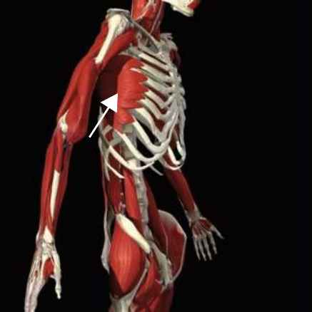
Lower trapezius
Origin
Spinous processes of T6–T12
Insertion
Spine of the scapula
Isolated Function
Concentric Action
Scapular depression
Integrated Function
Eccentric Action
Scapular elevation
Isometric Action
Stabilizes the scapula
Innervation
Cranial nerve XI, ventral rami C2–C4
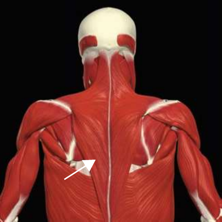
Middle trapezius
Origin
Spinous processes of T1–T5
Insertion
Acromion process of the scapula; superior aspect of the spine of the scapula
Isolated Function
Concentric Action
Scapular retraction
Integrated Function
Eccentric Action
Scapular protraction and elevation
Isometric Action
Stabilizes scapula
Innervation
Cranial nerve XI, ventral rami C2–C4
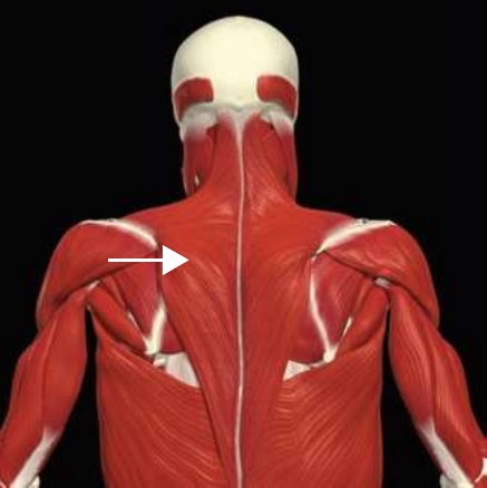
Upper trapezius
Origin
External occipital protuberance of the skull; spinous process of C7
Insertion
Lateral third of the clavicle; acromion process of the scapula
Isolated Function
Concentric Action
Cervical extension, lateral flexion and rotation; scapular elevation
Stabilizes the cervical spine and scapula; creates a stable base for the prime movers during scapular abduction and upward rotation
Innervation
Cranial nerve XI, ventral rami C2–C4
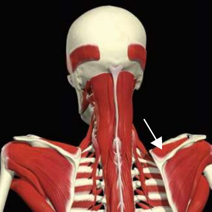
Levator scapulae
Origin
Transverse processes of C1–C4
Insertion
Superior vertebral border of the scapulae
Isolated Function
Concentric Action
Cervical extension, lateral flexion and ipsilateral rotation when the scapulae is anchored; assists in elevation and downward rotation of the scapulae
Integrated Function
Eccentric Action
Cervical flexion, contralateral cervical rotation, lateral flexion, scapular depression and upward rotation when the neck is stabilized
Isometric Action
Stabilizes the cervical spine and scapulae
Innervation
Ventral rami C3–C4, dorsal of subscapular nerve
Pectoralis major
Origin
Anterior surface of the clavicle; anterior surface of the sternum, cartilage of ribs 1–7
Insertion
Greater tubercle of the humerus
Isolated Function
Concentric Action
Shoulder flexion (clavicular fibers), horizontal adduction and internal rotation
Integrated Function
Eccentric Action
Shoulder extension, horizontal abduction and external rotation
Isometric Action
Stabilizes the shoulder girdle
Innervation
Medial and lateral pectoral nerve (C5–C7)
Pectoralis minor
Origin
Ribs 3–5
Insertion
Coracoid process of the scapula
Isolated Function
Concentric Action
Protracts the scapula
Integrated Function
Eccentric Action
Scapular retraction
Isometric Action
Stabilizes the shoulder girdle
Innervation
Medial pectoral nerve (C6–T1)
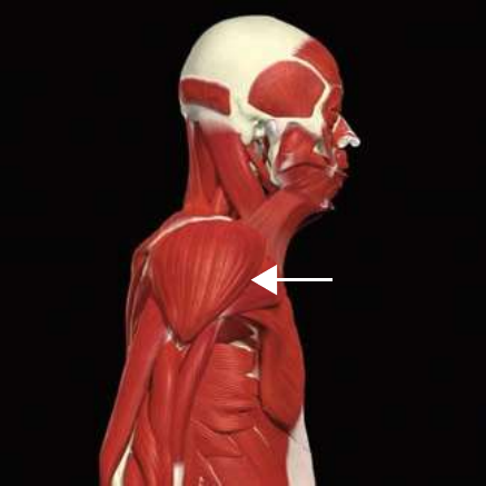
Anterior deltoid
Origin
Lateral third of the clavicle
Insertion
Deltoid tuberosity of the humerus
Isolated Function
Concentric Action
Shoulder flexion and internal rotation
Integrated Function
Eccentric Action
Shoulder extension and external rotation
Isometric Action
Stabilizes the shoulder girdle
Innervation
Axillary nerve (C5–C6)
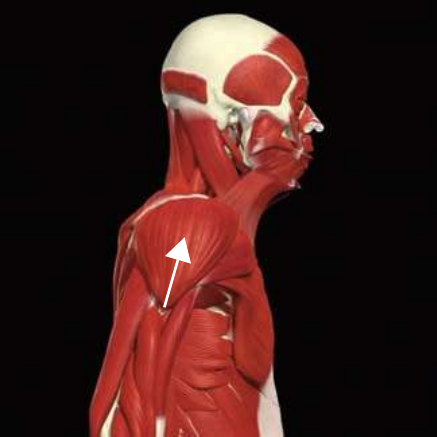
Medial deltoid
Origin
Acromion process of the scapula
Insertion
Deltoid tuberosity of the humerus
Isolated Function
Concentric Action
Shoulder abduction
Integrated Function
Eccentric Action
Shoulder adduction
Isometric Action
Stabilizes the shoulder girdle
Innervation
Axillary nerve (C5–C6)
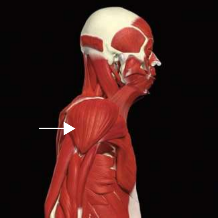
Posterior deltoid
Origin
Spine of the scapula
Insertion
Deltoid tuberosity of the humerus
Isolated Function
Concentric Action
Shoulder extension and external rotation
Integrated Function
Eccentric Action
Shoulder flexion and internal rotation
Isometric Action
Stabilizes the shoulder girdle
Innervation
Axillary nerve (C5–C6)
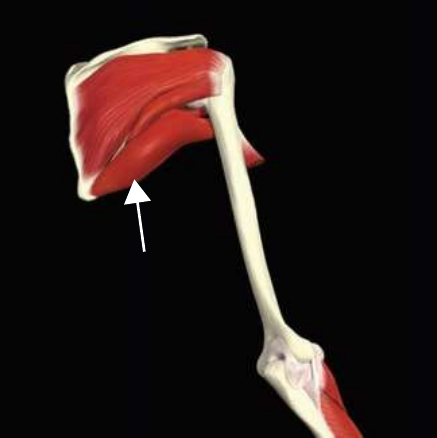
Teres minor
Origin
Lateral border of the scapula
Insertion
Greater tubercle of the humerus
Isolated Function
Concentric Action
Shoulder external rotation
Integrated Function
Eccentric Action
Shoulder internal rotation
Isometric Action
Stabilizes the shoulder girdle
Innervation
Axillary nerve (C5–C6)
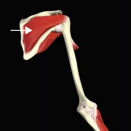
Infraspinatus
Origin
Infraspinous fossa of the scapula
Insertion
Middle facet of the greater tubercle of the humerus
Isolated Function
Concentric Action
Shoulder external rotation
Integrated Function
Eccentric Action
Shoulder internal rotation
Isometric Action
Stabilizes the shoulder girdle
Innervation
Suprascapular nerve (C5–C6)
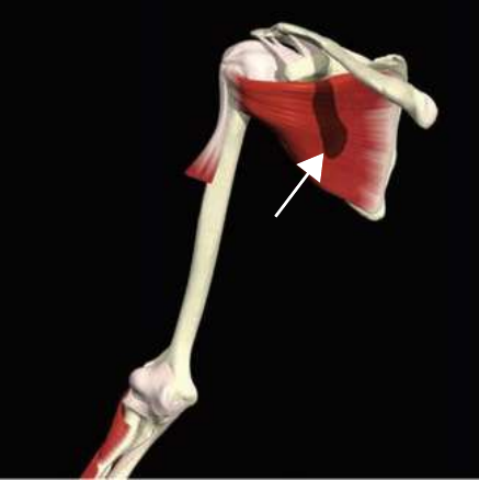
Subscapularis
Origin
Subscapular fossa of the scapula
Insertion
Lesser tubercle of the humerus
Isolated Function
Concentric Action
Shoulder internal rotation
Integrated Function
Eccentric Action
Shoulder external rotation
Isometric Action
Stabilizes the shoulder girdle
Innervation
Upper and lower subscapular nerve (C5–C6)
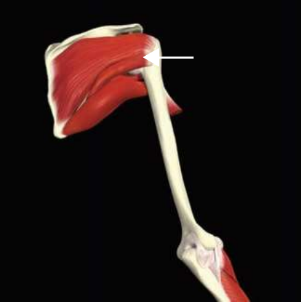
Supraspinatus
Origin
Supraspinous fossa of the scapula
Insertion
Superior facet of the greater tubercle of the humerus
Isolated Function
Concentric Action
Abduction of the arm
Integrated Function
Eccentric Action
Adduction of the arm
Isometric Action
Stabilizes the shoulder girdle
Innervation
Suprascapular nerve (C5–C6)
Teres major
Origin
Inferior angle of the scapula
Insertion
Lesser tubercle of the humerus
Isolated Function
Concentric Action
Shoulder internal rotation, adduction and extension
Integrated Function
Eccentric Action
Shoulder external rotation, abduction and flexion
Isometric Action
Stabilizes the shoulder girdle
Innervation
Lower subscapular nerve
ARM MUSCULATURE
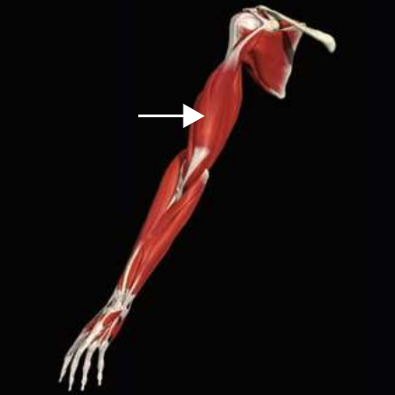
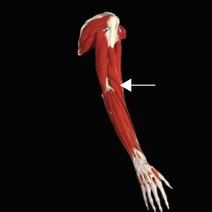
Biceps brachii
Origin
Short head: coracoid process; Long head: tubercle above the glenoid cavity on the humerus
Insertion
Radial tuberosity of the radius
Isolated Function
Concentric Action
Elbow flexion, supination of the radioulnar joint, shoulder flexion
Integrated Function
Eccentric Action
Elbow extension, pronation of the radioulnar joint, shoulder extension
Isometric Action
Stabilizes the elbow and shoulder girdle
Innervation
Musculocutaneous nerve
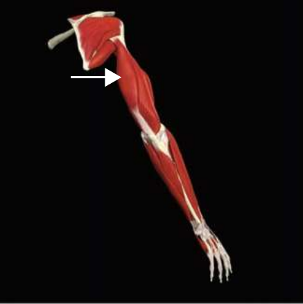
Triceps brachii
Origin
Long head: infraglenoid tubercle of the scapula; Short head: posterior humerus; Medial head: posterior humerus
Insertion
Olecranon process of the ulna
Isolated Function
Concentric Action
Elbow extension, shoulder extension
Integrated Function
Eccentric Action
Elbow flexion, shoulder flexion
Isometric Action
Stabilizes the elbow and shoulder girdle
Innervation
Radial nerve
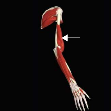
Brachialis
Origin
Humerus
Insertion
Coronoid process of the ulna
Isolated Function
Concentric Action
Flexes the elbow
Integrated Function
Eccentric Action
Elbow extension
Isometric Action
Stabilizes the elbow
Innervation
Musculocutaneous, radial nerve
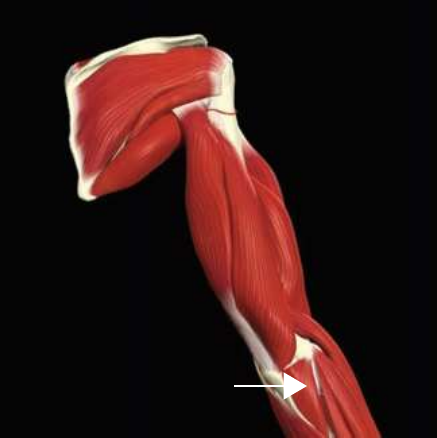
Anconeus
Origin
Lateral epicondyle of the humerus
Insertion
Olecranon process, posterior ulna
Isolated Function
Concentric Action
Extends the elbow
Integrated Function
Eccentric Action
Elbow flexion
Isometric Action
Stabilizes the elbow
Innervation
Radial nerve
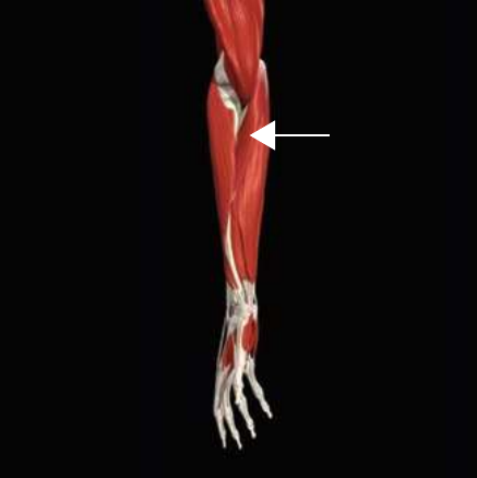
Brachioradialis
Origin
Lateral supracondylar ridge of the humerus
Insertion
Styloid process of the radius
Isolated Function
Concentric Action
Flexes the elbow
Integrated Function
Eccentric Action
Elbow extension
Isometric Action
Stabilizes the elbow
Innervation
Radial nerve
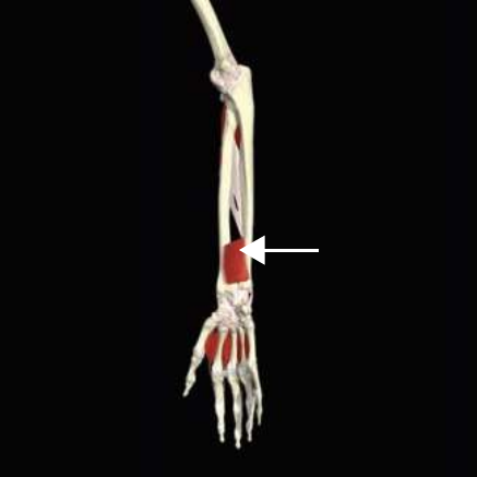
Pronator quadratus
Origin
Distal ulna
Insertion
Distal radius
Isolated Function
Concentric Action
Pronates the forearm
Integrated Function
Eccentric Action
Forearm supination
Isometric Action
Stabilizes the distal radioulnar joint
Innervation
Anterior interosseous nerve
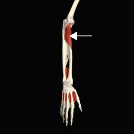
Pronator teres
Origin
Medial epicondyle of the humerus, coronoid process of the ulna
Insertion
Radius
Isolated Function
Concentric Action
Pronates the forearm
Integrated Function
Eccentric Action
Forearm supination
Isometric Action
Stabilizes the proximal radioulnar joint and elbow
Innervation
Median nerve
Supinator
Origin
Lateral epicondyle of the humerus
Insertion
Radius
Isolated Function
Concentric Action
Supinates the forearm
Integrated Function
Eccentric Action
Forearm pronation
Isometric Action
Stabilizes the proximal radioulnar joint and elbow
Innervation
Radial nerve
NECK MUSCULATURE
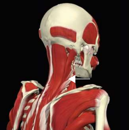
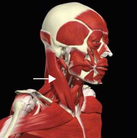
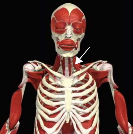
Sternocleidomastoid
Origin
Sternal head: top of the manubrium of the sternum; Clavicular head: medial one-third of the clavicle
Insertion
Mastoid process, lateral superior nuchal line of the occiput of the skull
Isolated Function
Concentric Action
Cervical flexion, rotation and lateral flexion
Integrated Function
Eccentric Action
Cervical extension, rotation and lateral flexion
Isometric Action
Stabilizes the cervical spine and acromioclavicular joint
Innervation
Cranial nerve XI
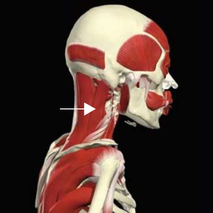
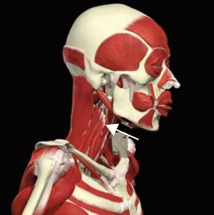
Scalenes
Origin
Transverse processes of C3–C7
Insertion
First and second ribs
Isolated Function
Concentric Action
Cervical flexion, rotation and lateral flexion; assists rib elevation during inhalation
Integrated Function
Eccentric Action
Cervical extension, rotation and lateral flexion
Isometric Action
Stabilizes the cervical spine
Innervation
Ventral rami (C3–C7)
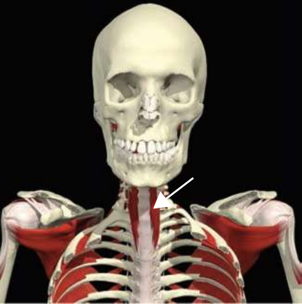
Longus colli
Origin
Anterior portion of T1–T3
Insertion
Anterior and lateral C1
Isolated Function
Concentric Action
Cervical flexion, lateral flexion and ipsilateral rotation
Integrated Function
Eccentric Action
Cervical extension, lateral flexion and contralateral rotation
Isometric Action
Stabilizes the cervical spine
Innervation
Ventral rami (C2–C8)
Longus capitus
Origin
Transverse processes of C3–C6
Insertion
Inferior occipital bone
Isolated Function
Concentric Action
Cervical flexion and lateral flexion
Integrated Function
Eccentric Action
Cervical extension
Isometric Action
Stabilizes the cervical spine
Innervation
Ventral rami (C1–C3)
Putting it togetherTable 2.4 — Muscle Synergies
Compound exercises recruit a whole team: a prime mover to drive the motion, synergists to assist, and a long list of stabilizers to keep every joint in the chain controlled. This is why "core" and lower‑leg muscles show up as stabilizers even in an upper‑body lift.
Table 2.4 visualized — even a "simple" squat coordinates prime movers, synergists, and stabilizers from the foot to the neck.
The skeletal levers and muscular forces only matter if the nervous system can organize them. Motor control is the study of posture and movements with the mechanisms available to control them. Motor learning looks at how movements are learned and retained for future use — proper practice and experience produce a permanent change in the ability to perform skilled movements. For that change to stick, feedback is essential.
Feedback: the use of sensory information and sensorimotor integration to help develop permanent neural representations of motor patterns for efficient movement. It comes in two broad forms — internal (sensory) feedback from proprioceptors, and external (augmented) feedback such as knowledge of results delivered after a movement.
Proprioception — the cumulative neural input from sensory receptors in the muscles, tendons, ligaments, and joints — is what lets the HMS sense joint position and adjust on the fly. Training that challenges proprioception and uses good feedback is how movement quality is rebuilt after dysfunction.
The one‑paragraph summary. Each component of the HMS — muscular (anatomy), skeletal (biomechanics), and nervous (motor behavior) — is interdependent. Movement is described in three planes (sagittal/frontal/transverse) around three axes, and almost no real motion is purely single‑plane. Every muscle has an isolated (concentric) function and integrated (eccentric and isometric) functions, and muscles work in four roles — agonist, antagonist, synergist, stabilizer — through length‑tension relationships, force‑couples, and bony levers to produce efficient, controlled human movement.